
Figure 2. Ca2+ influx through TRPV4 activates TMEM16F scramblase in BeWo cells.
(A) 20 nM GSK101 triggers Ca2+ influx and PS exposure (labeled by AnV) in wild-type (WT) BeWo cells. (B) 20 nM GSK101 induces Ca2+ influx, but fails to trigger PS exposure in TMEM16F knockout (KO) BeWo cells. Ca2+ dye (Calbryte 520, green) and fluorescently tagged AnV (AnV-CF594, red) were used to monitor the dynamics of intracellular Ca2+ and PS externalization, respectively. (C–D) Time course of GSK101-triggered Ca2+ influx (C) and PS exposure (D) in BeWo WT (n=5) and TMEM16F KO cells (n=5). AnV, Annexin V.
Figure 2—source data 1. Source data for Figure 2 and Figure 2—figure supplement 1B.
elife-78840-fig2-data1.xlsx (45.6KB, xlsx)
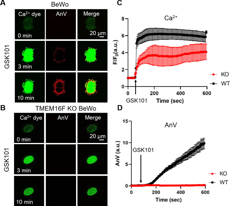
Figure 2—figure supplement 1. 20 nM GSK101 triggers Ca2+ increase and subsequent CaPLSase activities in primary human placental trophoblasts.

(A) Ca2+ dye (Calbryte 520) and fluorescently tagged AnV (AnV-CF594) were used to measure the dynamics of intracellular Ca2+ and PS externalization, respectively. All fluorescence images are the representatives of at least three biological replicates. (B) Time course of GSK101 triggered PS exposure in primary human placental trophoblasts. n=6. Error bar represents ± SEM. AnV, Annexin V.