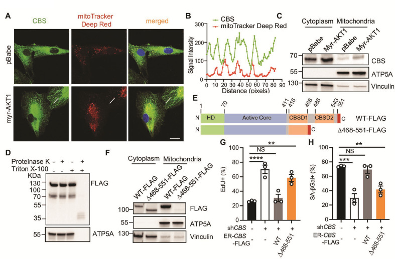
Figure 4. Cystathionine-β-synthase (CBS) mitochondrial localization is required for AKT-induced senescence (AIS) maintenance.
(A–B) BJ3 human skin fibroblasts expressing telomerase reverse transcriptase (BJ-TERT) cells were transduced with pBabe or myrAKT1. Immunofluorescent staining showing CBS (green) and mitochondria (red) on day 10 post-transduction. The representative images are from one of two independent experiments. Scale bar = 20 μm. (B) Quantification of signal intensities using ImageJ by applying a single ROI to two color channels in the same image and extracting the plot profile. (C) Western blot analysis of CBS expression in the cytoplasmic and mitochondrial fractions isolated from BJ-TERT cells transduced with pBabe or myrAKT1. ATP5A and vinculin serve as the markers of mitochondria and cytoplasm, respectively. (D) Western blot analysis of a protease protection assay using the mitochondrial fraction isolated from BJ-TERT cells expressing C-terminal FLAG-tagged CBS. ATP5A serves as a positive control. (E) Schematic of 4-OHT-inducible plasmids expressing FLAG-tagged wild type CBS (WT) or a C-terminal regulatory domain CBSD2 truncated CBS mutant. (F) Western blot analysis of CBS expression in the cytoplasmic and mitochondrial fractions isolated from BJ-TERT cells transduced with FLAG-tagged wild type (WT) or a truncated mutant CBS after 20 nM 4-OHT induction for 3 days. ATP5A and vinculin serve as the markers of mitochondria and cytoplasm, respectively. (C), (D), and (F) are representative of at least n=3 experiments. (G–H) BJ-TERT cells expressing doxycycline-inducible CBS shRNA#2 and 4-OHT-inducible CBS wide type or a truncated mutant were transduced with myr-AKT1, treated with doxycycline (1 μg/ml)±4 OHT (20 nM) on day 5 post-transduction and analyzed on day 12 post-transduction. The percentage of cells with positive staining for (G) EdU proliferation marker incorporation or (H) SA-βGal activity is expressed as mean ± SEM (n=3). One-way analysis of variance (ANOVA) with Holm-Šídák’s multiple comparisons was performed (NS, not significant; **p<0.01; ***p<0.001; ****p<0.0001).
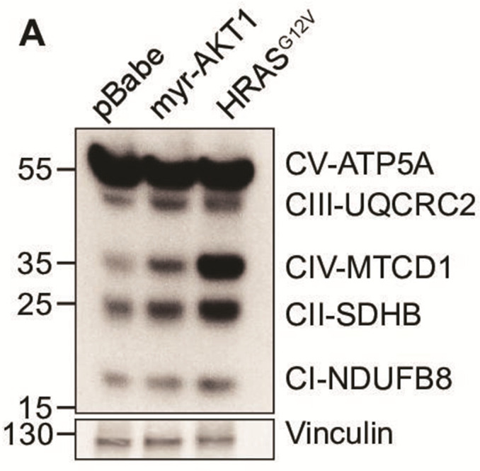
Figure 4—figure supplement 1. AKT-induced senescent cells showed increased expression of proteins involved in the mitochondrial electron transport chain.


