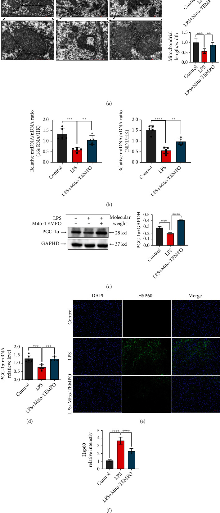
Figure 4.

Mito-TEMPO improves mitochondrial function in LPS-induced sepsis. (a) Representative transmission electron microscopy images of mitochondria in the liver of LPS-induced sepsis mice (scale bar = 2 μm, 1 μm). (b) Measurement of mtDNA copy numbers in the liver of mice. (c) The liver protein levels of PGC-1α as assessed by western blot. (d) The liver mRNA levels of PGC-1α as assessed by qPCR. (e, f) The proportion of HSP60-positive cells in the liver was determined by immunofluorescence (scale bar = 50 μm). n = 5 per group. Data are presented as the mean ± SD from at least three independent experiments. ∗P < 0.05, ∗∗P < 0.01, ∗∗∗P < 0.005, and ∗∗∗∗P < 0.0001.
