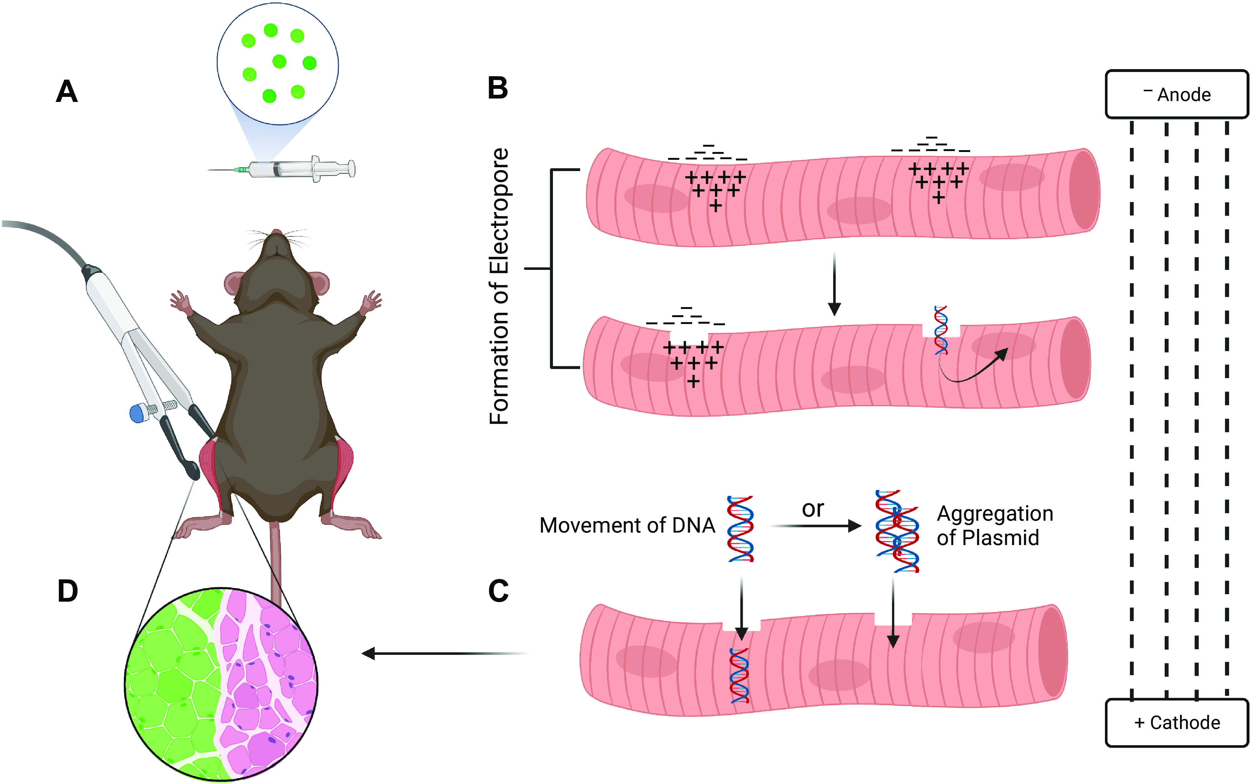
Figure 1.

Proposed mechanisms for the movement of plasmid DNA into electroporated muscle fibers. Generally, in vivo electroporation experiments allow for a within-animal study design to be used where one side of the animal is electroporated with an expression plasmid containing a gene of interest and the contralateral leg serves as the empty vector control. A: after plasmid injection, each leg is exposed to electrical pulses through an electroporation generator via electrodes placed on the targeted hindlimb muscle. B: application of electrical pulses to the muscle generates positive and negative charge on either side of the membrane. In turn, this charge imbalance leads to the formation of hydrophobic pore defects enabling plasmid DNA to enter the myofiber. C: movement of plasmid DNA into the myofiber is proposed to occur either through movement toward the anode electrophoretically or by localizing with other DNA molecules at the cell membrane and moving together into the myofiber as a complex without electrophoretic forces being required. D: these proposed mechanisms contribute to the successful introduction of the plasmid DNA into the cell resulting in a transfected myofiber [depicted as green fluorescent protein (GFP) positive]. Adapted from McMahon and Wells (27). Created with Biorender.com.
