Figure 1.

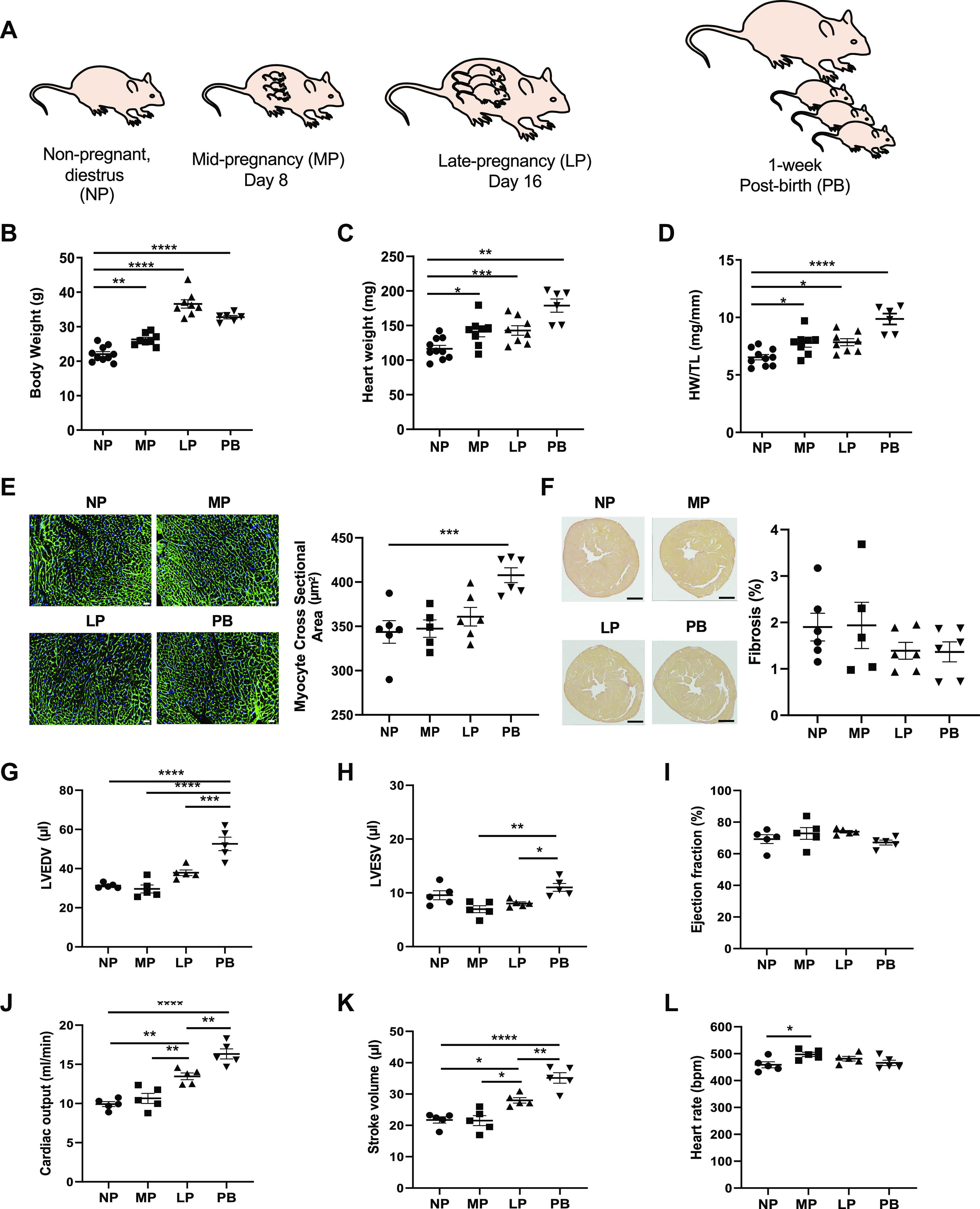
Pregnancy and postpartum period are associated with structural and functional remodeling of the maternal heart. A: schematic of study design showing the time points analyzed, which include nonpregnant, diestrus (NP), midpregnant (MP; day 8 of pregnancy), late pregnant (LP; day 16 of pregnancy), and 1-wk postbirth with lactation (PB). B–D: gravimetric measurements of body weight (B), heart weight (C), and heart weight-to-tibia length ratio (HW/TL; D) in NP, MP, LP, and PB female mice (n = 6–10 per group). E: representative images of wheat germ agglutinin and quantification of myocyte cross section. Scale bars, 200 μm. F: representative images of Sirius Red staining and quantification of % fibrosis from NP, MP, LP, and PB female mice (n = 6 per group). G–L: echocardiographic measurements of left ventricular end-diastolic volume (LVEDV; G), left ventricular end-systolic volume (LVESV; H), ejection fraction (I), cardiac output (J), stroke volume (K), and heart rate (L) from NP, MP, LP, and PB female mice (n = 5 per group). The respective n numbers in this figure correspond to 3 parallel groups of mice. *P < 0.05, **P < 0.01, ***P < 0.001, ****P < 0.0001, ANOVA with Tukey’s post hoc test.
