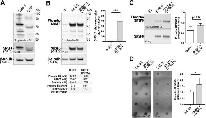
Fig. 3.
SRSF6 phosphorylation in iPSC cardiomyocytes overexpressing DYRK1A. A Evaluation of the anti-phosphoepitope SR antibody in total protein extracts from AC16 cardiomyocytes treated with calf intestine alkaline phosphatase (CIAP). B Detection of phospho-SRSF6 and SRSF6 in iPSC cardiomyocytes transfected with empty vector (EV), SRSF6-HA expression construct (SRSF6) or co-transfected with DYRK1A and SRSF6-HA (SRSF6 + DYRK1A). β-Tubulin was assayed as loading control. Lower panel: densitometric analysis. Upper right panel: relative DYRK1A mRNA fold expression. (A.U.: arbitrary units). C Detection of phosphorylated SRSF6 in immunoprecipitated samples from iPSC cardiomyocytes. Right panel: densitometric analysis. Horizontal bars show the mean ± SD from determinations performed in quintuplicate. D Dot-blot analysis of phosphorylated SRSF6 in immunoprecipitated samples. Right panel: densitometric analysis. Horizontal bars show the mean ± SD from determinations performed in quintuplicate. ***P < 0.001, *P < 0.05, Student’s t test

