Figure 3.
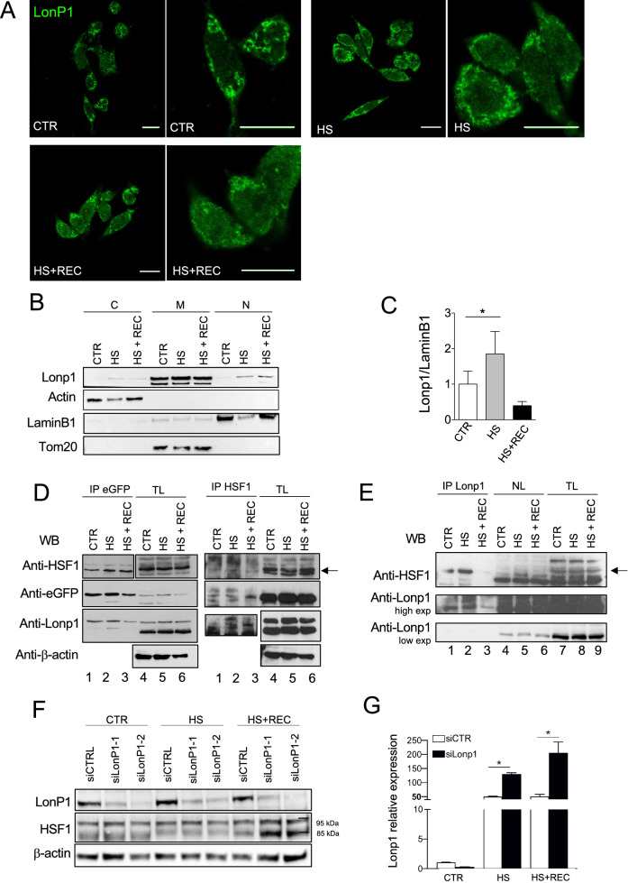
Lonp1 relocates in the nucleus in response to heat shock. (A) Representative confocal microscopy images of SW620 cells after immunostaining with anti-Lonp1 antibody. Cells were kept at 37 °C (CTRL), heat-shocked (HS) at 42 °C and then left to recover (HS + REC). Bars: 10 mm. (B) Representative immunoblot showing Lonp1 expression in cytosolic, mitochondrial and nuclear fractions from SW620 cells maintained at 37 °C (CTR), at 42 °C for 3 h to induce heat shock (HS) and kept at 42 °C for 3 h and then left for 1 h at 37 °C to recover (HS + REC). β-actin is the cytosolic fraction loading control, TOM20 is the mitochondrial fraction loading control and lamin B1 is the nuclear fraction loading control. Blots were cut prior to hybridisation with antibodies during blotting. (C) Histogram representing the relative levels of Lonp1 in nuclear fractions, obtained from three independent experiments. Data are shown as mean SD. *p < 0.05. (D) Representative immunoprecipitation experiment showing the interaction between Lonp1-eGFP and HSF1 in SW620 cells after heat-shock (HS) and then left 3 h at 37 °C to recover (HS + REC). Left panels: lysates were immunoprecipitated with anti-eGFP and immunoblotted with anti-HSF1. Western blot on total lysate (TL) is also shown. Blots were cut prior to hybridisation with antibodies during blotting. Right panels: lysates were immunoprecipitated with anti-HSF1 and immunoblotted with anti-eGFP. Blots were cut prior to hybridisation with antibodies during blotting. (E) Representative immunoprecipitation experiment showing the interaction between endogeneous Lonp1 and HSF1 in nuclear lysates from SW620 cells after heat-shock (HS) and then left 3 h at 37 °C to recover (HS + REC). Nuclear lysates were immunoprecipitated with anti-Lonp1 and immunoblotted with anti-HSF1. Western blots on nuclear lysates (NL) and total lysates (NL) are also shown. Blots were cut prior to hybridisation with antibodies during blotting. (F) Representative immunoblot showing the quantification of HSF1 in SW620 cells transfected with scramble small-interfering RNAs (siCTRL) and cells transfected with small-interfering RNAs against Lonp1 (siLonp1-1 and siLonp1-2), after HS and HS + REC. Blots were cut prior to hybridisation with antibodies during blotting. (G) Quantification of the mRNA levels of HSP70 in SW620 cells transfected with scramble small-interfering RNAs (siCTRL) and cells transfected with small-interfering RNAs against Lonp1 (siLonp1), after HS and HS + REC. *P < 0.05.

