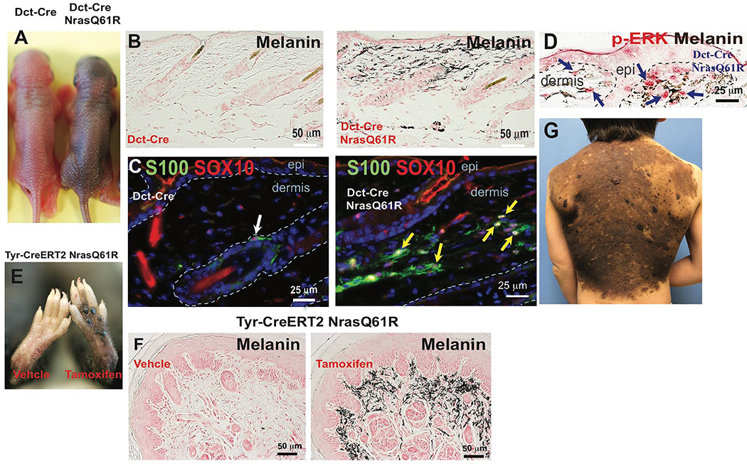
Figure 1. Melanocyte-specific NrasQ61R mutant mice recapitulates histologic and molecular features of human giant congenital melanocytic nevi.
(A) Features of melanocytic nevi in 3-day-old Dct-Cre NrasQ61R. (B) Melanin of adult Dct-Cre NrasQ61R mice was detected by Fontana-Masson staining. (C-D) Immunostains. Paraffin sections were immunostained for S100 (C, green), SOX10 (C, red), or p-ERK (D, red). (E) Features of melanocytic nevi in tamoxifen-induced Tyr-CreERT2 NrasQ61R mice. (F) Melanin in tamoxifen-induced Tyr-CreERT2 NrasQ61R mice was detected by Fontana-Masson staining. (G) The clinical appearance of a human giant congenital nevus is shown in. White arrow in (C) indicates normal signal in hair follicle. Yellow arrows in (C) indicate ectopic signals in the dermis of nevus skin. Blue arrows in (D) indicate positive signals. See also supplementary Figures 1 and 2.

