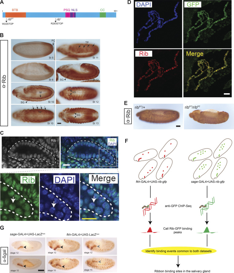
Figure 2.
Strategy for identifying transcriptional targets of Rib in the SG. (A) Rib protein diagram with the N-terminal Bric à Brac, Tramtrack, Broad (BTB) domain, the pipsqueak (PSQ) DNA-binding domain, the bipartite nuclear localization sequence (NLS), and a predicted coiled coil (CC) domain. Compound heterozygotes from the null alleles of rib—ribP7 and rib1—were used throughout this study. (B) Rib is broadly expressed during embryogenesis. M, mesoderm; PC, pole cells; TP, tracheal primordia. Scale bar: 50 µm. (C) Rib is nuclear. Top: White outline marks the SG. Bottom: Rib staining is reduced in the DAPI-intense areas (nucleoli; arrow). Scale bar: 10 µm. (D) Rib-GFP, used for tissue-specific ChIP-seq analysis, localizes to SG polytene chromosomes of L3 larvae; note the one-to-one correspondence of bands detected with αGFP and αRib antisera. (Images from sage-GAL4 > UAS-rib-GFP.) Scale bar: 10 µm. (E) Validation of the αRib guinea pig antiserum used for the experiments in Figs. 2 D and 7 B. ribP7 sibling heterozygotes and homozygotes were stained in the same tube to confirm the nuclear localization of Rib antigen in heterozygotes and its loss in homozygotes. Nonspecific background (diffuse, nonnuclear staining) from the HRP reaction with the secondary antibody was observed in all embryos. Scale bar: 50 µm. (F) Strategy for ChIP-seq using fkh-GAL4 or sage-GAL4 to drive UAS-Rib-GFP. Tissue expression patterns of Rib-GFP in fkh-GAL4 > UAS-rib-GFP embryos and sage-GAL4 > UAS-rib-GFP embryos are illustrated in red (SG, hemocytes, and mesoderm) and green (SG and midgut), respectively. The SG is the only tissue with Rib-GFP expression from both the drivers; thus, the intersection of binding peaks from both driver datasets represent high-confidence SG-specific binding. (G) Expression of SG drivers used in the Rib-GFP ChIP-seq are indicated with nuclear βgal during early (stage 12) and late (stage 15) tubulogenesis. For the fkh-GAL4 driver, a different focal plane (right images) captures expression in mesodermally derived cells. Arrowheads mark the SG. Scale bar: 50 µm.

