Fig. 6.
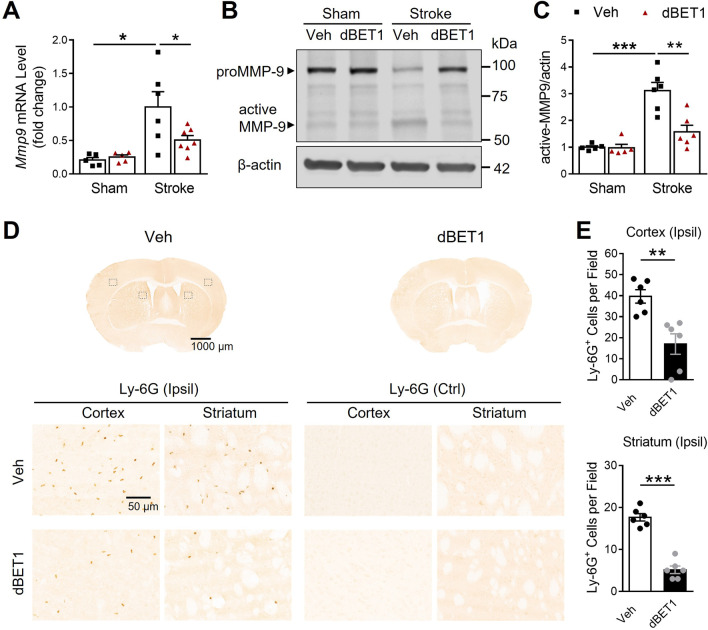
dBET1 attenuates ischemia-induced MMP-9 increase and neutrophil infiltration. A Matrix metalloproteinase-9 (MMP-9) mRNA level of the ischemic and sham cortex was measured by real-time PCR. Stroke led to a dramatic increase in MMP-9 mRNA level, which was significantly reduced by dBET1. n = 5 per sham group, n = 6 stroke veh group, n = 7 stroke dBET1 group. *P < 0.05. B Representative immunoblot for MMP-9 level in homogenates from the ischemic cortex and sham controls. C dBET1 significantly reduced active MMP-9 protein level in the ipsilateral (Ipsil) cerebral cortex compared to the veh group. n = 5 per sham group, n = 6 per stroke group. **P < 0.01, ***P < 0.001. D Representative images of staining for Ly-6G, a specific marker of neutrophils, in the post-stroke mouse brain sections at 48 h after tMCAO, showing the neutrophilic infiltration; and open squares in the left image indicate the peri-infarct areas of the ipsilateral cortex and striatum, as well as the contralateral (Ctrl) side, used for micrographic examination. No Ly-6G stain signal is detected in the contralateral side of both groups. Accumulated Ly-6G staining is widely distributed in the peri-infarct area, which is much lower in the dBET1 group. per field, 200 µm × 200 µm square. E Quantifications of Ly-6G in D showed that the dBET1 group exhibits reduced expression level compared to veh group (> 50% in ischemic cortex and > 70% in ischemic striatum. n = 6 per group. **P < 0.01, ***P < 0.001

