Fig. 8.
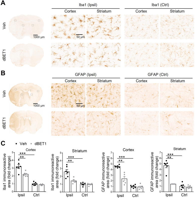
dBET1 attenuates ischemia-induced reactive gliosis. Representative images of Iba1 positive microglia (A) and GFAP positive astrocyte (B) in the cortex and striatum of mice at 48 h after tMCAO; and open squares in the left image indicate the peri-infarct areas of the ipsilateral (Ipsil) cortex and striatum, as well as the contralateral (Ctrl) side, used for micrographic examination. C Quantifications of the areas of Iba1 and GFAP signals in A and B respectively. Acute stroke evoked reactive gliosis, the response of glial cells to ischemic insults, in the peri-infarct area, in microglia (characterized by hypertrophic soma with thickened and retracted processes) and astrocytes (characterized by hypertrophic somas and highly stained processes). dBET1 treatment remarkably attenuated the deteriorative progression of reactive gliosis in microglia and astrocytes compared to veh controls. In addition, no obvious difference was detected in both markers in the contralateral cortex and striatum regions. n = 6 per group. *P < 0.05, **P < 0.01. Veh, vehicle

