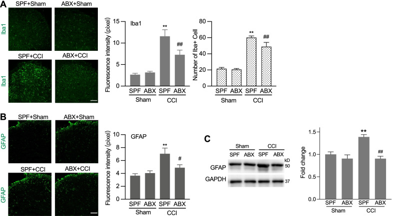
Fig. 3.
ABX treatment alleviated CCI-induced activation of microglia and astrocyte in the spinal cord of mice. A Representative images of the Iba-1 microglia immunostaining (scale bar: 100 µm) and the quantification of the fluorescence intensity (upper) and cell numbers (lower) of the Iba1 immunostained cells. One-way ANOVA with Dunnett multiple comparison test **p < 0.01 versus SPF, ##p < 0.01 versus SPF + CCI (15 slides from 4 SPF sham mice, 15 slides from 4 ABX sham mice, 12 slides from 4 SPF + CCI mice, and 11 slides from 3 ABX + CCI mice). B Representative images of the GFAP astrocyte immunostaining (scale bar: 100 µm) and the quantification of the fluorescence intensity the GFAP immunostaining cells. One-way ANOVA with Dunnett multiple comparison test **p < 0.01 versus SPF, #p < 0.05 versus SPF + CCI (48 slides from 4 SPF mice, 51 slides from 4 ABX mice, 28 slides from 4 SPF + CCI mice, and 28 slides from 3 ABX + CCI mice). C Representative image of bands and statistical summary of the western blot analysis showing that CCI-induced GFAP increase was reversed by ABX treatment. n = 3 in each group. **p < 0.01 versus SPF, ##p < 0.01 versus SPF + CCI. Tissues were taken from SPF and ABX-treated mice at day 7 after CCI or sham operation (A–C)

