Fig. 5.
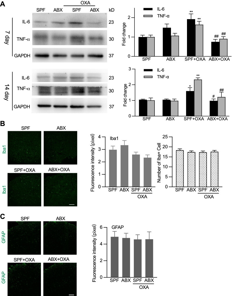
Differential effects of ABX treatment on chemotherapy-induced cytokine production in DRG and glia cell activation in the spinal cord. A Western blotting analysis showing that ABX treatment suppressed the expression of IL-6 and TNF-α in DRG in day 7 and 14 after OXA treatment. Left: representative Western blot bands. Right: statistical summary. One-way ANOVA with Tukey multiple comparison test *p < 0.05, **p < 0.01 SPF + OXA versus SPF; #p < 0.05, ##p < 0.01 ABX + OXA versus SPF + OXA (on day 7: IL-6, n = 6 in each group; TNF-α, n = 7 in ABX, n = 6 in other groups, and on day 14: n = 3 in each group). B, C Both OXA and ABX treatment did not alter the activation of microglial cells and astrocytes in the spinal cord. Left: representative images of immunostaining of Iba1 and GFAP. Scale bar: 100 µm. Right: quantification of the fluorescence intensity and cell numbers of Iba1 immunostaining. Tissues were taken 7 days after OXA treatment (B and C). In B, 15 slides from 4 mice in SPF, 16 slides from 4 mice in ABX, 16 slides from 4 mice in SPF + OXA, 17 slides from 4 mice in ABX + OXA. In C, 6 slides from 4 mice in each group

