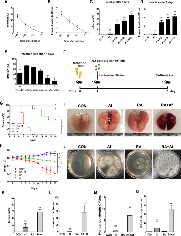
Fig. 1.
The establishment of a mouse model of post-radiation Aspergillus fumigatus infection. a Infection rates in C57 mice after intratracheal challenge by A. fumigatus conidia (5 × 10⁷/ml) were calculated at 24 h, 48 h, 72 h, and 7 Days. b Mice lung tissue homogenates were filtered and plated to measure log10 of colony-forming units (CFU). c, d The infection rate and fungal burden of different radiation doses observed after 7 days. e Rate of A. fumigatus infection when administered at different time points after irradiation. f Experiment process. g, h Survival status and changes in body weight in each group. i Representative images of lung tissues. j Lung tissue homogenates of each group were inoculated on Sabouraud dextrose agar, and k infection rates were calculated for each group. l, m The number of colonies and log10 of CFU in lung tissue homogenates. n Real-time polymerase chain reaction (PCR) analysis results for A. fumigatus and 18S rRNA levels in the lungs. Data are shown as the mean ± standard deviation (n = 8). *P < 0.05, **P < 0.01 versus the control group

