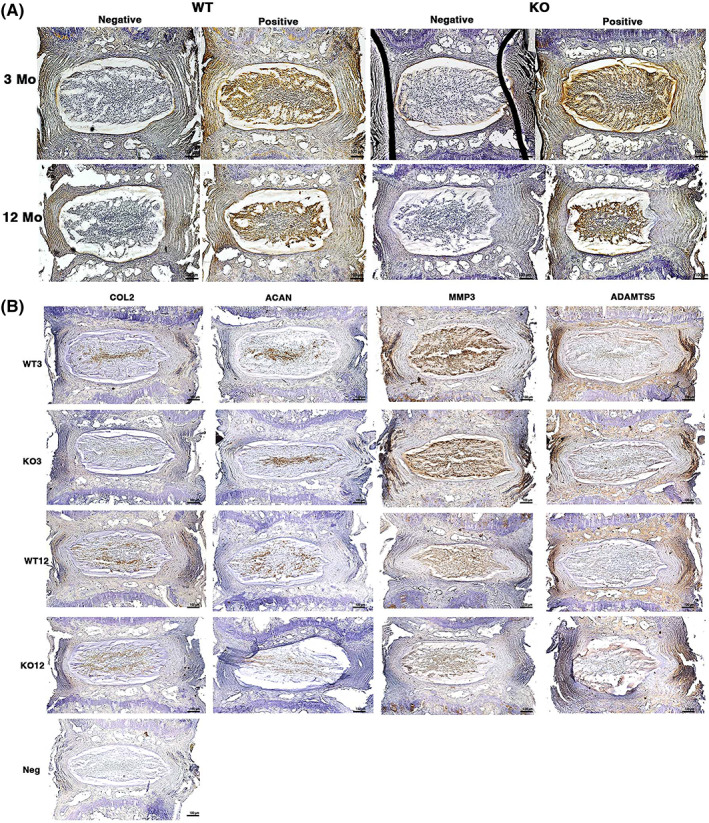
FIGURE 4.

(A) Immunohistochemistry of IL‐1β and matrix proteins in uninjured caudal IVDs. IVDs underwent immunohistochemical staining with anti‐IL‐1β antibody and biotinylated secondary antibody, with HRP‐DAB (dark brown color) visualization. Intense IL‐1β staining was seen in the NP in most samples, with no difference between WT and IL‐1Ra−/−. (B) Immunohistochemistry of matrix anabolic proteins (Col2 and ACAN) and catabolic enzymes (MMP‐3 and ADAMTS‐5). Despite some differences in distribution between NP and AF for MMP‐3 (more in NP) and ADAMTS‐5 (more in AF), there were no differences between groups, even though there were gene expression differences
