FIG 2.

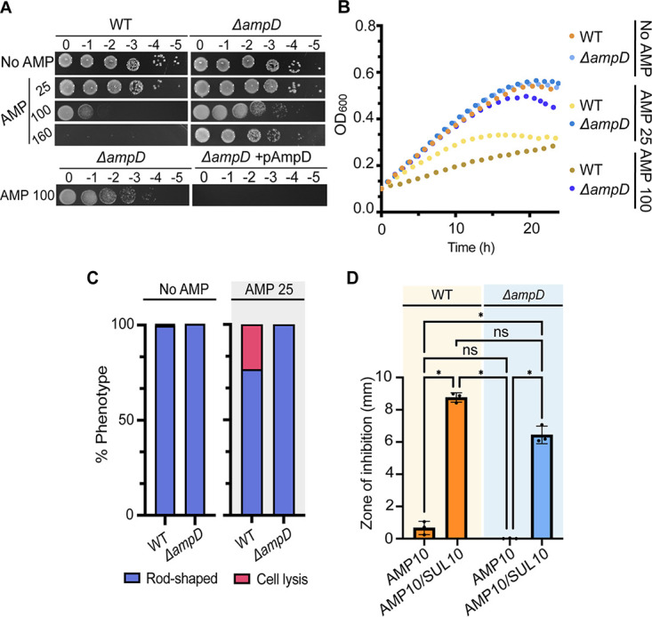
Loss of AmpD results in constitutive β-lactamase activity and elevated ampicillin resistance. (A) Ampicillin susceptibility assay performed by spotting dilutions. Briefly, exponential cultures were serially diluted and spotted on solid medium containing no ampicillin (No AMP) or ampicillin at 25, 100, or 160 μg/mL (AMP 25, AMP 100, or AMP 160, respectively) and incubated at 28°C for ~40 h before imaging. Plates used to demonstrate complementation of ΔampD (ΔampD + pAmpD) included 1 μM IPTG to induce expression of plasmid-encoded AmpD. (B) Growth of A. tumefaciens WT and ΔampD in the absence (No AMP) and presence of various concentrations of ampicillin (AMP 25 or AMP 100) for 24 h (n = 1; 2 replicates). (C) Quantitative analysis of phase-contrast microscopy of exponentially growing strains treated with ampicillin at 25 μg/mL (AMP 25). The percent phenotype was calculated by counting the number of cells displaying one of the indicated phenotypes (1 cell = 1 phenotype) and dividing it by the total number of cells for each strain. (D) Disk susceptibility assay performed on a lawn of indicated strains grown on LB plates for 24 h at 28°C. AMP 10, disk containing 10 μg/mL ampicillin; AMP 10/SUL 10, disk containing 10 μg/mL ampicillin and 10 μg/mL sulbactam, a broad-spectrum β-lactamase inhibitor. Data represent the mean (±SD) of three independent experiments. ****, P < 0.0001; ***, P < 0.001; **, P < 0.01; *, P < 0.1; ns, not significant.
