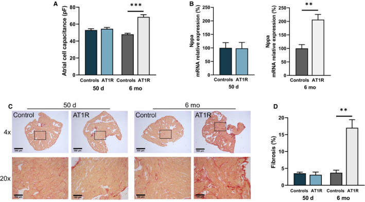
Figure 1. Structural remodeling is only present in angiotensin II type 1 receptor (AT1R) mice at 6 months.

A, Cell capacitance measurements in left atrial myocytes from 50‐day (controls, n=60; AT1R, n=62 [P=0.1918]) and 6‐month (controls, n=29; AT1R, n=20 [***P<0.0001]) mice indicate that cellular hypertrophy is only observed in 6‐month AT1R mice. B, The relative messenger RNA (mRNA) expression of Nppa was unchanged between AT1R and control mice at 50 days (n=4; P=0.9517) but was increased in 6‐month AT1R mice compared with controls (controls, n=3; AT1R, n=4 [**P=0.0050]). C, An example of Picrosirius red staining of paraffin‐embedded left atrial sections of 50‐day and 6‐month controls and AT1R mice at 4× and 20× magnification. Collagen deposits are stained in red and myocardium in yellow. D, Interstitial fibrosis signal quantification (%) of 50‐day and 6‐month mice (n=3 per group) shows an increase only in 6‐month AT1R mice (50 days, P=0.5685; 6 months, **P=0.0030). Statistical analysis of Figures 1, 2, 3, 4, 5, 6, 7, 8 was performed using 2‐tailed Student t test. Statistical significance interpretation: *P<0.05; **P<0.01; ***P<0.0001.
