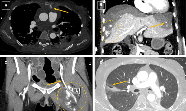
Figure 2. Illustrative radiographic examples of cardiopulmonary resuscitation complications.

(A and C) Anterior mediastinal hematoma (arrow, A) and left thigh medial compartment hematoma (circle) associated with a femoral artery catheter (arrow, C), both exhibiting active extravasation in a patient with disseminated intravascular coagulation treated with massive transfusion. (B) Extensive subcapsular liver hematoma with multiple foci of active extravasation (circle, arrow) and several lacerations treated conservatively because of a poor prognosis with multiple transfusions and eventual autotamponade of bleeding. (D) Right lower lobe pulmonary laceration (arrow) associated with bilateral rib and sternal fractures in a patient with hereditary pheochromocytoma/paraganglioma syndrome who underwent prolonged resuscitation.
