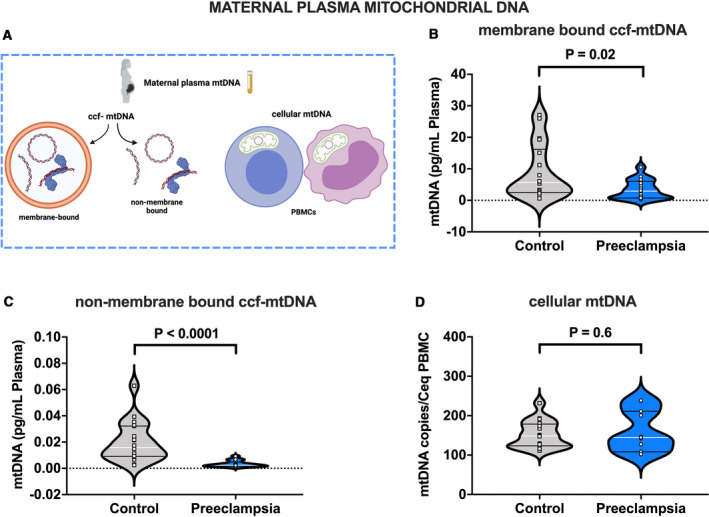
Figure 1. Mitochondrial DNA in cell‐free and cellular forms in maternal plasma from pregnancies with preeclampsia and healthy controls.

A, Biological forms of mtDNA assessed in this study, (B) ccf‐mtDNA concentrations (pg/mL) in plasma isolated using lysis buffer (membrane bound ccf‐mtDNA), (C) ccf‐mtDNA concentrations (pg/mL) in plasma isolated without lysis buffer (non‐membrane bound ccf‐mtDNA), (D) cellular mtDNA copies per estimated number of peripheral blood mononuclear cells (PBMCs), Student’s t‐test (B and D), Mann‐Whitney U test (C). B through C, n=18 controls, n=17 preeclampsia; (D) n=16 controls, n=11 preeclampsia. All values presented as median (white line), IQR (black lines). ccf‐mtDNA indicates circulating cell‐free mitochondrial DNA; Ceq, cell equivalent; and PBMC, peripheral blood mononuclear cell. Open squares=gestational age‐matched control; open circles=third trimester pregnancies with preeclampsia. A, was created with Biorender.com.
