
Figure 6. Regions of highly repetitive LRA after adenosine infusion identified at 20% cutoff threshold (in pink/purple) occur at regions of LRA at baseline at lower frequency in patient 10 (top images) in contrast with patient 8 (lower panel) in whom adenosine promotes LRA in regions widely distributed throughout the chamber with little correlation to those zones seen at baseline.
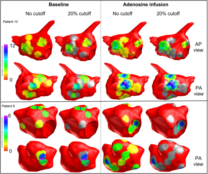
Mitral annulus is outlined in black, areas of excluded occurrences are shown in grey. AP indicates anterior posterior; LRA, localized rotational activation; and PA, posterior anterior.