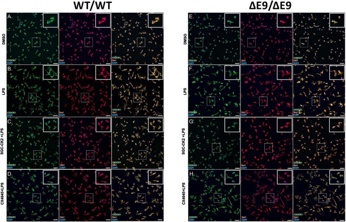
FIGURE 4.
Characterization of DMSO, LPS and drug treated MGLs derived from WT/WT and ΔE9/ΔE9 (FAD) iPSCs. WT (A–D) and FAD (E–H) MGLs were stimulated with LPS (B,F) and simultaneously treated with either SGC-CK2-1 (C,G) or CX-4945 (D,H) for 24 h and immunocytochemistry was performed. In all conditions, MGLs robustly express microglia specific markers, CX3CR1 and Iba1. Scale bar = 50 μm.

