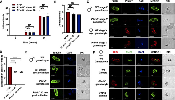
FIG 2.
Pfarid– parasites grow normally as asexual parasites and undergo gametocytogenesis but fail to form microgametes. (A) Parasite growth rate for WT PfNF54 and Pfarid– (clone 4E and 6A) was measured over two erythrocytic cycles using Giemsa-stained smears. Data were averaged from three biological replicates and are presented as the mean ± standard deviation (SD). (B) Gametocytemias for WT PfNF54 and Pfarid– (clones 4E and 6A) parasites were measured on day 15 using Giemsa-stained smears. Data were averaged from three biological replicates and are presented as the mean ± SD. (C) IFAs performed for day 15 WT or Pfarid– stage V gametocytes (clone 4E) using α-P230p (green), a stage V male-specific marker, and Pfg377 (red), a marker for female gametocytes. (D) The number of exflagellation centers (vigorous flagellar beating of microgametes in clusters of RBCs) per field at 15 min postactivation. Data were averaged from three biological replicates and are presented as the mean ± SD. (E) IFAs performed on WT or Pfarid– (clone 4E) gametocytes activated for 20 min in vitro using α-tubulin II (green), a male-specific marker. α-Tubulin II staining showed male gametes emerging from an exflagellating male gametocyte in WT PfNF54. The Pfarid– gametocytes were defective for male gametocyte exflagellation. ND, not detected. (F) IFAs performed on WT PfNF54 and Pfarid– (clone 4E) gametocytes activated for 20 min in vitro using Pfs25 (green), a marker for female gametes, and PfUIS4, marker for the parasitophorous vacuole membrane. Female gametes did not show any defect in egress from the infected RBC. NS, not significant; ND, not detected.

