FIG 3.
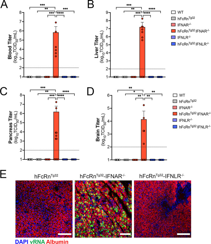
Type I IFNs control echovirus dissemination from the GI tract. Seven-day-old pups were orally inoculated with 106 PFU of E5, and at 3 dpi, animals were sacrificed for viral titration and histology. (A to D) Viral titers in the blood (A), liver (B), pancreas (C), and brain (D). In all panels, titers are shown as log10 TCID50 per milliliter, with the limit of detection indicated by a dotted line. Data are shown as means ± standard deviations, with individual animals shown as each data point. (E) Hybridization chain reaction (HCR) RNA fluorescence in situ hybridization (RNA-FISH) from liver sections of hFcRnTg32, hFcRnTg32-IFNAR−/−, or hFcRnTg32-IFNLR−/− neonatal mice at 3 dpi using probes against the E5 genome (green) and albumin (red). DAPI-stained nuclei are shown in blue. Bars, 100 μm. In panels A to D, data are shown with significance determined by a Kruskal-Wallis test with Dunn’s test for multiple comparisons (*, P < 0.05; **, P < 0.005; ***, P < 0.0005; ****, P < 0.0001).

