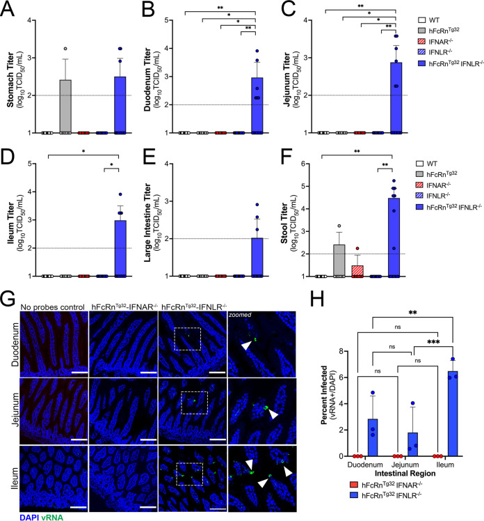
FIG 4.
Type III IFNs restrict persistent echovirus infection in the GI epithelium. Seven-day-old neonatal mice were orally inoculated with 106 PFU of E5, and at 7 dpi, animals were sacrificed for viral titration and tissue collection. (A to F), Viral titers in the stomach (A), duodenum (B), jejunum (C), ileum (D), large intestine (E), and stool (F). In all panels, titers are shown as log10 TCID50 per milliliter, with the limit of detection indicated by a dotted line. Data are shown as means ± standard deviations, with individual animals shown as each data point. Significance was determined using a Kruskal-Wallis test with Dunn’s test for multiple comparisons (*, P < 0.05; **, P < 0.005). (G) At 3 dpi, animals were sacrificed, and the entire GI tract was removed and Swiss rolled, followed by histological sectioning. HCR of hFcRnTg32-IFNAR−/− or hFcRnTg32-IFNLR−/− pups at 3 dpi was performed using probes against the E5 genome (green) and DAPI (blue). Bars, 100 μm. Zoomed-in views of specific regions of the hFcRnTg32-IFNLR−/− images are shown on the right. (H) Quantification of three independent tile scans using confocal microscopy of each region of the small intestine based on the number of villi that were positive for vRNA using the cell count function in FIJI. Data are shown as a percentage of vRNA-positive villi over total villi per tile scan. Three independent tile scans were quantified (for an average of 144 villi in the duodenum, 224 villi in the jejunum, and 164 villi in the ileum). Significance was determined by two-way ANOVA with Šídák’s multiple-comparison tests (*, P < 0.05; **, P < 0.005; ***, P < 0.0005; ****, P < 0.0001).

