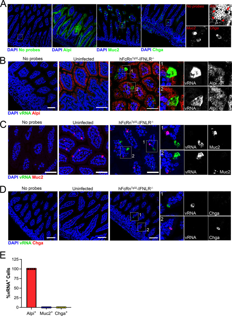
FIG 5.
In vivo replication of echoviruses is specific for enterocytes. (A) Hybridization chain reaction (HCR) RNA-FISH of uninfected small intestine sections using specific probes against Alpi, Muc2, or Chga (in green), as indicated at the bottom. DAPI-stained nuclei are shown in blue. A no-probe-containing control is shown on the left. In all panels, the white box is shown zoomed in (magnification, approximately ×6) at the right using the probes indicated in red. Red arrows in the Alpi section denote goblet cells based on morphology that were not positive for Alpi, as expected. (B to D) Seven-day-old hFcRnTg32-IFNLR−/− neonatal mice were orally inoculated with 106 PFU of E5; at 3 dpi, animals were sacrificed; and the entire small intestine was removed and Swiss rolled for subsequent histological sectioning. Shown are representative images of ileum tissue using probes for E5 (green in all panels) and either Alpi (B), Muc2 (C), or Chga (D) (red in all panels). DAPI-stained nuclei are shown in blue. In all panels, white boxes denote zoomed-in areas shown on the right, which include black-and-white images, as indicated. Bars, 50 μm. (E) Quantification of confocal images was performed using FIJI and expressed as the total percentage of vRNA-positive cells that colocalized with Alpi (in red), Muc2 (in blue), or Chga (in green). Note that there was no colocalization between vRNA and either Muc2 or Chga.

