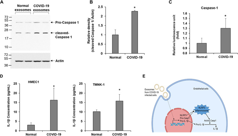
FIG 2.
Exosomes isolated from COVID-19 patients activate caspase-1 and induce 1L-1β secretion. (A) HMEC-1 cells were exposed to exosomes from normal and COVID-19 patients for 48 h, and cell lysates were subjected to Western blot analysis for caspase-1 using a specific antibody. The membrane was reprobed for actin as an internal control. (B) The quantitative presentation of band intensities using Image J software is shown on the right. (C) Caspase-1 activity was measured in exosomes exposed to HMEC-1 culture medium using the Caspase-Glo 1 inflammasome assay reagent. Luminescence was read after 3 h of incubation with the reagent, and results are presented as relative luminescence unit. (D) HMEC-1 or TMNK-1 cells were exposed to exosomes from normal and COVID-19 patients for 48 h, and IL-1β from culture medium was assayed using the ELISA Max deluxe set human IL-1β kit. Relative absorbance was measured at 450 nm. The concentration of IL-1β in the medium was calculated from a standard curve. The small bar indicates standard error (*, P < 0.05). (E) The schematic presentation shows exosomes secreted from SARS-CoV-2-infected cells that trigger NLRP3, pro-caspase-1 (Casp1), and pro-IL-1β transcription resulting in the activation of Casp1 followed by IL-1β via the NLRP3 inflammasome in endothelial cells.

