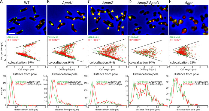
FIG 2.
Polar organizers are required for polar localization of the origins. (A to E) Visualization of ori1 (green) and ori2 (red) in WT (AtWX263) (A) and indicated ΔpodJ (AtWX307) (B) ΔpopZ (AtWX303) (C), ΔpopZ ΔpodJ (AtWX305) (D), and Δgpr (AtW309) (E) mutants. ori1 and ori2 are labeled using GFP-ParB1 and RFP-RepBCh2 expressed from a single pSRKKm-based plasmid. (Top) Cropped fluorescence microscopy images. (Middle) Plots of focus positions. (Bottom) Plots of distance of foci from the nearest pole. Scale bar represents 2 μm. Yellow carets point to the foci that are distant from the cell pole. Colocalization was defined as a pair of green and red foci that are within an interfocal distance of fewer than 6 pixels. Detailed image analysis can be found in Fig. S2 and Materials and Methods.

