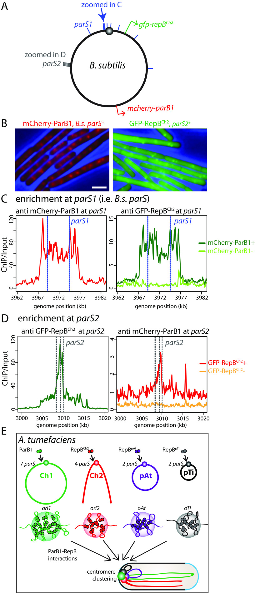
FIG 4.
ParB1 and RepBCh2 interact in B. subtilis. (A) Schematic of the B. subtilis chromosome. Blue bars indicate the position of the nine B. subtilis parS sites, which have the same consensus sequence as A. tumefaciens parS1 sites. Blue arrow points to the two parS1 sites shown in panel C. Gray bars indicate the cluster of parS2 sites also shown in panel D. Red and green arrows indicate the locations from which mcherry-parB1 and gfp-repBCh2 are expressed. (B) Expressing mCherry-ParB1 (left) and GFP-RepBCh2 (right) in the presence of parS1 and parS2 leads to fluorescence focus formation in B. subtilis. (C, left) At parS1 sites (blue dotted lines), mCherry-ParB1 had high enrichment. (Right) GFP-RepBCh2 had low enrichment when mCherry-ParB1 was present, and no enrichment was seen when mCherry-ParB1 was absent. (D) At parS2 sites (gray dotted lines), GFP-RepBCh2 had high enrichment (left); mCherry-ParB1 has low enrichment when GFP-RepBCh2 was present and no enrichment when GFP-RepBCh2 was absent (right). (E) Schematic model of ori clustering. Centromeric ParB1 and RepB protein dimers bind to their parS sites (11) near the replication origins of the four replicons and spread to nearby regions to form nucleoprotein complexes. The three RepB nucleoprotein complexes interact with the ParB1 nucleoprotein complex, leading to the clustering of the four origins/centromeres.

