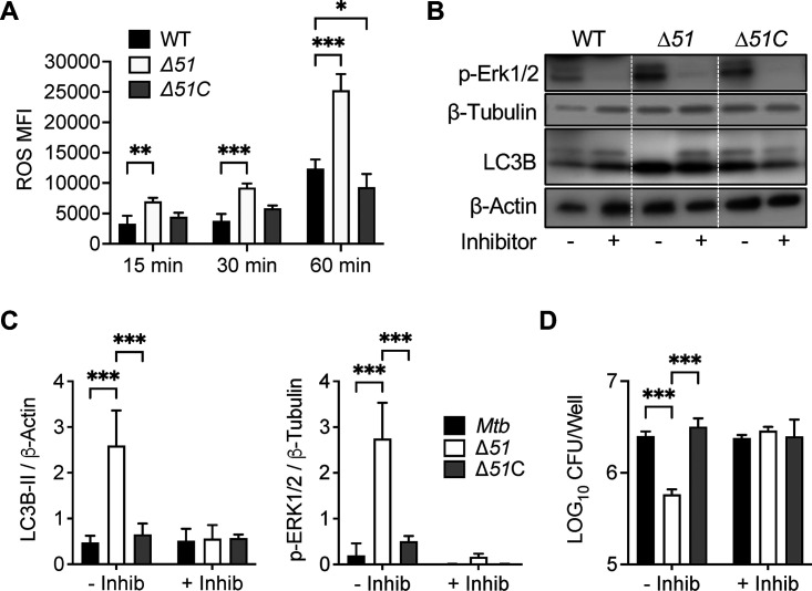
FIG 2.
Role of ROS generation and MAP kinases in autophagy induction by Δ51. (A) RAW 264.7 macrophages treated with the oxidative stress reagent CellROX were infected with WT, Δ51, and Δ51C at an MOI of 10. At 15 min, 30 min, and 60 min, cells were collected and acquired by flow cytometry. (B) Immunoblot of phosphorylated ERK1/2 and LC3B accumulation in RAW 264.7 macrophages infected with WT, Δ51, and Δ51C at an MOI of 10, with or without ERK1/2 inhibitor (5 μM, FR180204) 24 h postinfection. A representative blot of three independent assays is shown. (C) The densitometric summary analysis was calculated by p-ERK1/2 density normalized to β-tubulin density and LC3B-II density to β-actin. (D) WT, Δ51, and Δ51C Mtb survival was determined in RAW 264.7 macrophages (MOI, 10) with or without ERK1/2 inhibitor (5 μM) at 24 h postinfection. All graphs represent one of three independent experiments, data are means and standard deviations (SD). Significance was calculated by two-way ANOVA corrected by Dunnett's test for multiple comparisons. *, P < 0.05; ***, P < 0.001.

