FIG 4.
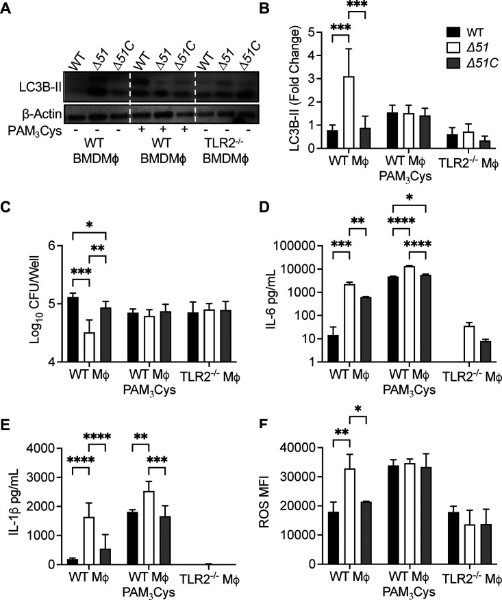
Mtb PPE51 inhibits TLR2 signaling. (A) Immunoblot of LC3B accumulation in WT BMDM with or without 100 ng/mL Pam3CSK4 and TLR2−/− BMDM infected with WT, Δ51, and Δ51C at an MOI of 10 at 24 h postinfection. Representative blot of three independent assays shown. (B) The densitometric summary analysis was calculated by LC3B-II density normalized to β-actin density, and then the fold change ratio was calculated relative to the uninfected control for each assay. (C) WT, Δ51, and Δ51C survival was determined in WT BMDM with or without 100 ng/mL Pam3CSK4 and TLR2−/− BMDM infected (MOI 10) at 24 h postinfection. (D and E) Cytokine concentration in WT BMDM with or without 100 ng/mL Pam3CSK4 and TLR2−/− BMDM culture supernatant 24 h postinfection. (F) ROS accumulation in WT BMDM with or without 100 ng/mL Pam3CSK4 and TLR2−/− BMDM 24 h postinfection. Macrophages were treated with CellROX 24 h postinfection, and ROS accumulation was determined by flow cytometry. The means and SD of representatives from three (B and C) or two (D to F) independent assays are shown. Significance was calculated by two-way ANOVA corrected by Dunnett's test for multiple comparisons (B to F). *, P < 0.05; **, P < 0.01; ***, P < 0.001; ****, P < 0.0001.

