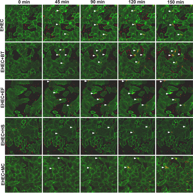
FIG 2.
Time-lapse microscopy of Lifeact::GFP-expressing HeLa cells infected with mCherry-expressing EHEC and commensals strains. A representative field of each infection condition was imaged for 2.5 h, and images taken every 5 min. White arrowheads indicate clusters of pedestal-forming bacteria. Original magnification: 63×.

