FIG 3.

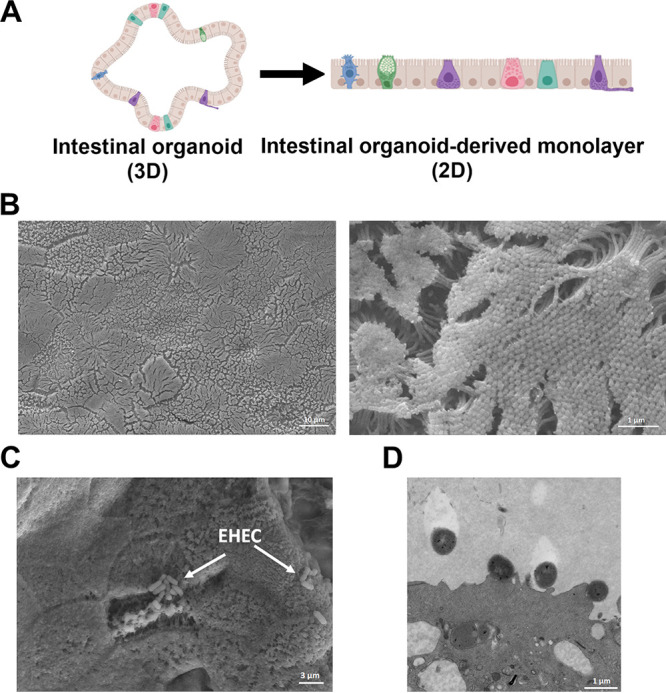
Human colonoids as a model for EHEC infection. (A) A schematic of 3D and 2D (monolayer) human colonoids. (B) Scanning electron microscope (SEM) images of uninfected colonoids (mock controls) showing the monolayer surface (low magnification, left panel) and highlighting the brush border and microvilli (high magnification, right panel). (C) SEM images of EHEC-infected colonoid monolayers after 6 h. White arrows point to clusters of attached EHEC. (D) Transmission electron microscope (TEM) images of infected colonoid monolayers showing the presence of actin pedestals formed by EHEC.
