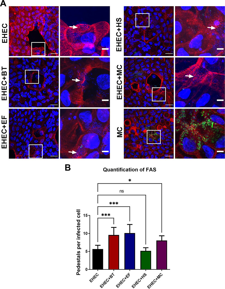
FIG 4.
Gut commensals enhance EHEC attachment to human colonoids. (A) FAS assay to detect pedestal formation on colonoid monolayers infected with EHEC and GFP-expressing commensal strains for 6 h. Actin and EHEC/DNA were stained with Alexa Fluor 568-phalloidin (red) and DAPI (blue), respectively. White arrows indicate sites of EHEC attachment and actin pedestal formation. Original magnification: 63×. Scale bar: 10 μm (left panels), 5 μm (right panels). (B) Quantification of FAS showing the number of pedestals per infected cells enumerated in multiple fields (n = 4). Error bars represent the means ± SD, and statistical significance was determined by one-way ANOVA followed by a post hoc Tukey test. *, P < 0.05; ***, P < 0.001; ns, not significant.

