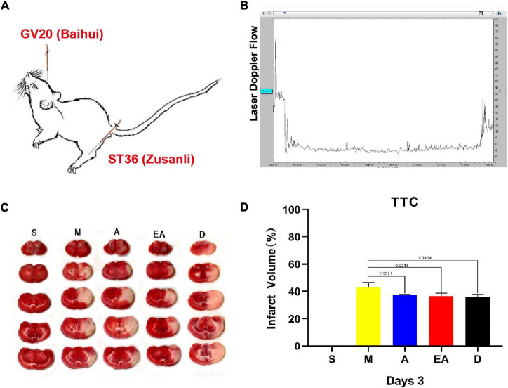
FIGURE 1.
Establishment of the rat middle cerebral artery occlusion (MCAO) model. (A) Detailed acupoint locations used for acupuncture and electroacupuncture. (B) Laser Doppler flux measured over the lateral parietal cortex in the core of the ischemic region of MCAO rats. (C) Representative images of 2,3,5-triphenyltetrazolium chloride (TTC) staining in rat brain slices from all groups (n = 3). (D) Quantification of infarct volumes in the whole hemisphere after 90 min of MCAO in rats. Data are presented as the mean percentage of the entire ischemic hemisphere ± SD.

