
Figure 5.
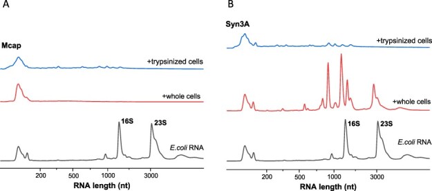
RNA degradation induced by surface RNases of (A) Mcap and (B) Syn3A cells. The control sample (bottom data line in panels A and B) shows intact RNA isolated from E. coli lysate. Addition of mycoplasma cells (middle data line in both panels SA and B) and trypsinized mycoplasma cells (top data line in both panels SA and B), caused E. coli rRNA to degrade into fragments <1500 nt. Compared to Mcap, Syn3A cells presented less active surface RNases.