
Figure 1.
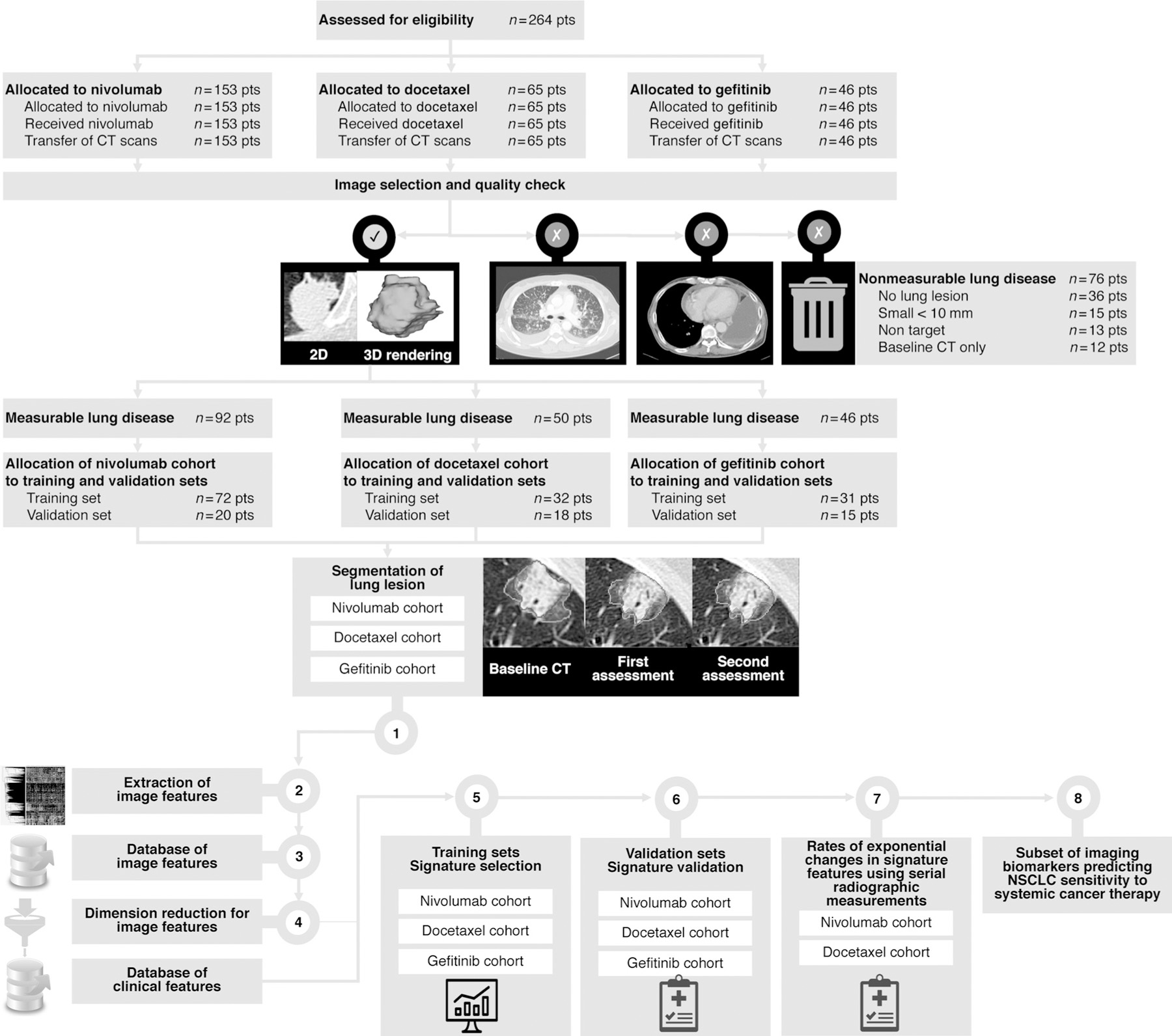
Disposition of study patients. Patients could be excluded for multiple reasons. The withdrawal boxes show the number of patients excluded at each step. CT scans acquired at sites are transferred to our academic core. Image selection and quality check using a computer-aided algorithm designed by machine learning. Step 1. Segmentation of the largest measurable lung tumor on CT scan by an expert radiologist at baseline in all patient (inclusion criteria), as well as all available radiographics measurement. Steps 2–3. Tumor imaging phenotype in each patient based on imaging features extraction in the largest measurable segmented lung lesion (1,160 imaging features characterizing changes between baseline and first CT assessment). Step 4. Dimension reduction using machine learning. Identification of reproducible, nonredundant, and informative candidate imaging features for model building. Step 5. Signature building in the training set to enhance strategic decision-making and predict treatment sensitivity. Step 6. Signature validation. Step 7. Transfer of the signature features for evaluation of g and d values using serial radiographic measurements. Step 8. A subset of imaging biomarker is identified.