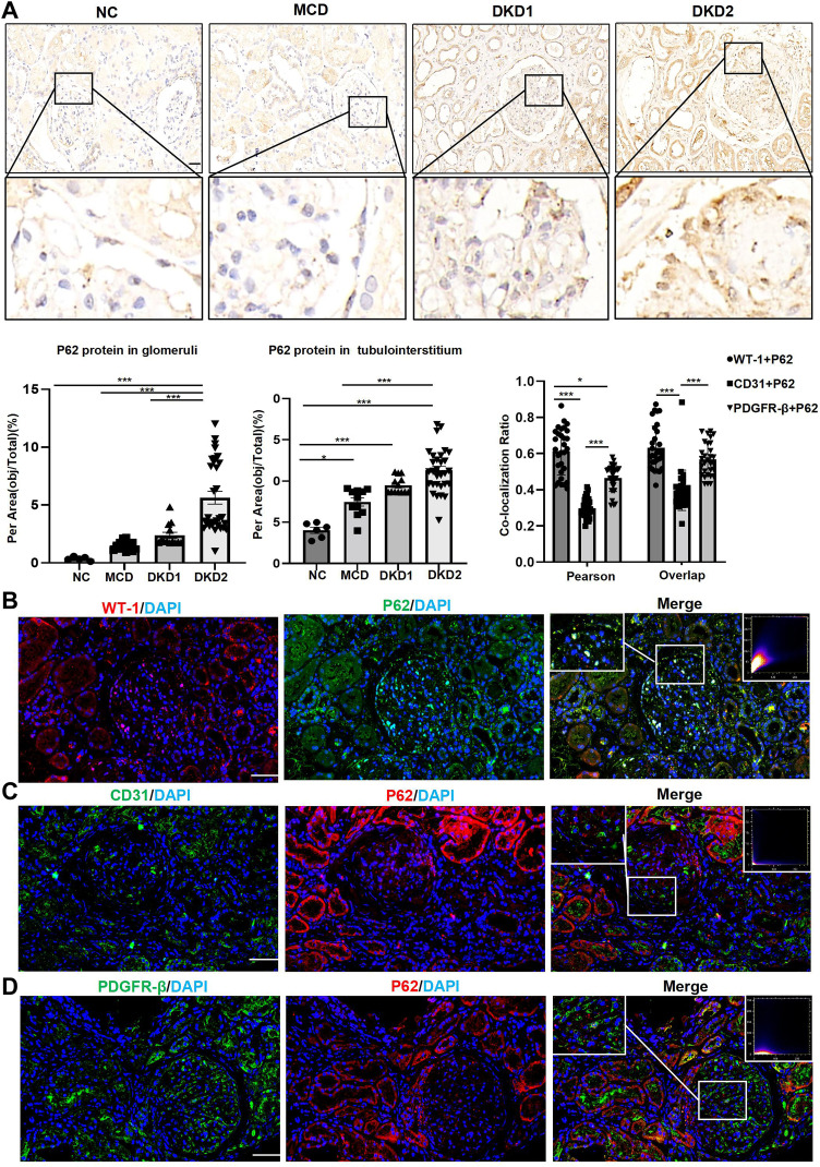
Figure 3.
The expressions of the P62 protein in human renal tissues were significantly increased in the DKD group patients with macroalbuminuria, mainly in the podocytes. (A) IHC staining of P62 protein in human renal tissues; (B) double immunofluorescence staining of P62 and WT-1 in human renal tissue of DKD2 group. (C) Double immunofluorescence staining of P62 and CD31 in human renal tissue of DKD2 group. (D) Double immunofluorescence staining of P62 and PDGFR-β in human renal tissue of DKD2 group. Data are expressed as mean ± standard deviation (SEM). Scale bar = 50 μm; *p < 0.05, ***p < 0.001.

