. 2022 Jun 28;11:e77365. doi: 10.7554/eLife.77365

Figure 6. Functional domain mapping of the microtubule nucleation and aster formation activity of calmodulin-regulated spectrin-associated protein 2 (CAMSAP2).

(A) Microtubule nucleation and aster formation activities of CAMSAP2 deletion constructs evaluated by the results of 10 µM tubulin with 1 µM CAMSAP2. The number of ‘+’ symbols indicates the strength of the activity (++++, strongest; +, weakest; ND, not detected). Size exclusion chromatography and SDS-PAGE of GFP fused constructs are available in Figure 6—figure supplement 1. CH, calponin-homology domain; MBD, microtubule-binding domain; CC, coiled-coil domain; CKK, C-terminal domain common to CAMSAP1 and two other mammalian proteins, KIAA1078 and KIAA1543. (B) Microtubule growth from CAMSAP2 condensates composed of full-length and deletion constructs. In vitro reconstitution was performed as described in Figure 4I. (C) Fluorescent intensity of CAMSAP2 condensates at 0 s. ND means that the fluorescent intensity of condensates could not be measured because GFP-CKK did not induce any condensates. Ordinary one-way ANOVA followed by Tukey’s multiple comparisons test. ****, p<0.0001. n=100 condensates from three independent preparations. (D) Negative stain EM images of polymerization by 10 µM tubulin with 1 µM GFP-CAMSAP2-FL or 1 µM CAMSAP2 mutants during 10 min of incubation at 37°C. The results for tubulin alone and GFP-CKK are available in Figure 6—figure supplement 3. Representative EM images are shown from at least three independent assays.

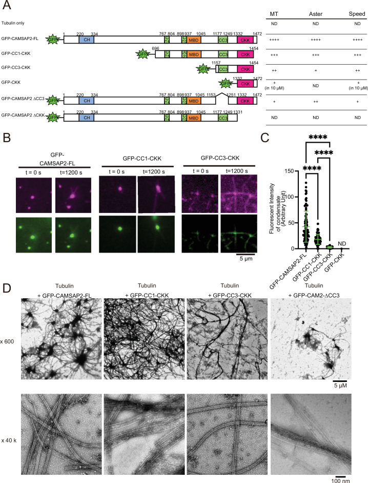
Figure 6—source data 1. Quantifications of fluorescent intensity of calmodulin-regulated spectrin-associated protein 2 condensates at 0 s.

Figure 6.

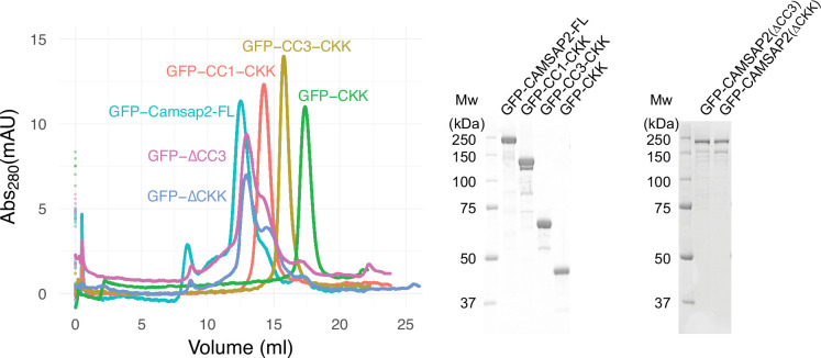
Figure 6—figure supplement 1. GFP fused calmodulin-regulated spectrin-associated protein 2 (CAMSAP2) constructs used in this study.

Figure 6—figure supplement 1.

Size exclusion chromatography of GFP-CAMSAP2-FL (turquoise green), GFP-CC1-CKK (orange), GFP-CC3-CKK (yellow), GFP-CAMSAP2 ∆CC3 (magenta), GFP-CAMSAP2 ∆CKK (blue), and GFP-CKK (green). The peaks of each sample were subjected to SDS-PAGE.
Figure 6—figure supplement 1—source data 1. SDS-PAGE gel of calmodulin-regulated spectrin-associated protein 2 deletion constructs.
Figure 6—figure supplement 1—source data 2. SDS-PAGE gel of calmodulin-regulated spectrin-associated protein 2 deletion constructs.
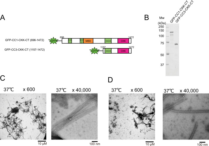
Figure 6—figure supplement 2. Functional domain mapping of the microtubule nucleation and aster formation activity of calmodulin-regulated spectrin-associated protein 2 (CAMSAP2) deletion mutants.

Figure 6—figure supplement 2.

(A) Schematic representation of the GFP fused CAMSAP2 deletion constructs. CH, calponin-homology domain; MBD, microtubule-binding domain; CC, coiled-coil domain; CKK, C-terminal domain common to CAMSAP1 and two other mammalian proteins, KIAA1078 and KIAA1543. (B) SDS-PAGE of the CAMSAP2 deletion constructs. (C) Negative stain electron microscopy (EM) micrographs of 10 µM tubulin polymerized with 1 µM of GFP-CC1-CKK-CT after incubation on ice for 30 min and 37°C for 10 min. (D) Negative stain EM micrographs of 10 µM tubulin polymerized with 1 µM of GFP-CC3-CKK-CT after incubation on ice for 30 min and 37°C for 10 min.
Figure 6—figure supplement 2—source data 1. SDS-PAGE gel of calmodulin-regulated spectrin-associated protein 2 deletion constructs.
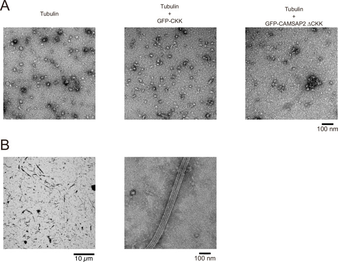
Figure 6—figure supplement 3. Functional domain mapping of calmodulin-regulated spectrin-associated protein 2 (CAMSAP2) analysed by negative stain electron microscopy (EM).

Figure 6—figure supplement 3.

(A) Ten-micromolar tubulin was incubated alone (left), or with 1 µM GFP-CKK (middle), or with 1 µM GFP-CAMSAP2 ∆CKK (right) at 37°C for 10 min and analysed by negative stain EM. Oligomerization of tubulin was observed in these three conditions, albeit no microtubule formation was observed. (B) Ten-micromolar tubulin with 10 µM GFP-CKK was incubated at 37°C for 10 min and analysed by negative stain EM. Notably, the microtubules were fully decorated with the GFP-CKK.