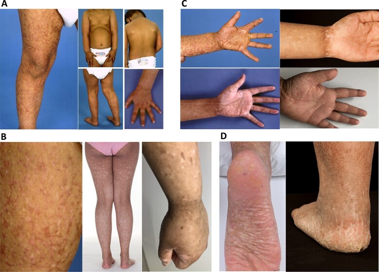
Figure 2.

Clinical synopsis. (A) Patient 1, aged 2.5 years, showing the distribution skin changes. (B) Morphology of skin changes in (left to right) Patient 1 at 2.5 and 14 years and Patient 4 at 21 years. (C) Palms of Patient 1 (left), aged 2.5 and 7 years, and of Patient 4 (right) aged 11 and 21 years, showing reticulate pigmentation, progressive scaling and peripheral keratoses. (D) Feet of Patient 1 (left) aged 14 years and of Patient 4 (right) aged 11 years.
