
Figure 2.
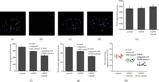
Distribution of motor neurons from the zygomatic branch and the buccal branch at the facial nerve nucleus. The blue color shows motor neurons of the buccal branch of the facial nerve stained with FG. The red color shows DIL-stained zygomatic motor neurons of the facial nerve. The claret color demonstrates the double-labeled neurons. (a) Normal distribution of motor neurons from the zygomatic branch and the buccal branch motor neurons; (b) normal control group (nerve conduit filled with water); (c) testing group with the nerve conduit filled with GDNF solution; (d) testing group with the nerve conduit filled with GDNF microcapsule; (e) summary of total regenerated neurons. Total regenerated neurons = total regenerated neurons in the zygomatic branch + total regenerated neurons in the buccal branch–double − labeled neurons; (f) summary of total incorrectly regenerated neurons. Total incorrectly regenerated neurons = incorrectly regenerated neurons in the zygomatic branch + incorrectly regenerated neurons in the buccal branch + double − labeled neurons; (g) summary of double-labeled neurons; (h) summary of the total incorrect regeneration ratio. Total incorrect regeneration ratio = total incorrectly regenerated neurons/total regenerated neurons in the zygomatic branch and buccal branches.