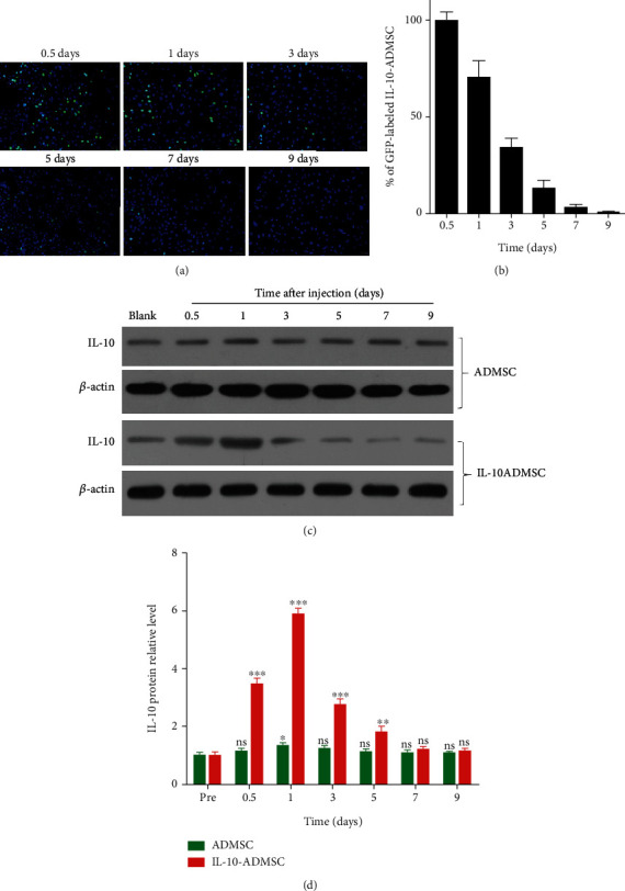
Figure 5.

IL-10-modified ADMSC underwent extensive apoptosis and survived for a short time after injection. (a) In the wound, the locally injected IL-10-ADMSC gradually reduced which was evaluated by observing green fluorescence, and representative tissue fluorescence image was displayed; (b) the GFP-labeled IL-10-ADMSC in the wound after different time of local injection of IL-10-ADMSC were statistically compared; (c–d) after different time of local injection of IL-10-ADMSC, Western blot was used to detect the expression of IL-10 in the wound. Representative protein bands were displayed and statistically compared protein band gray value. Scale bar: 10 μm. Data was expressed as (SD ± mean), and each analysis was repeated at least 3 times independently; P value was calculated by Student's t test; ns was P > 0.05, ∗ was P < 0.05, ∗∗ was P < 0.01, and ∗∗∗ was P < 0.001 vs. ADMSC group.
