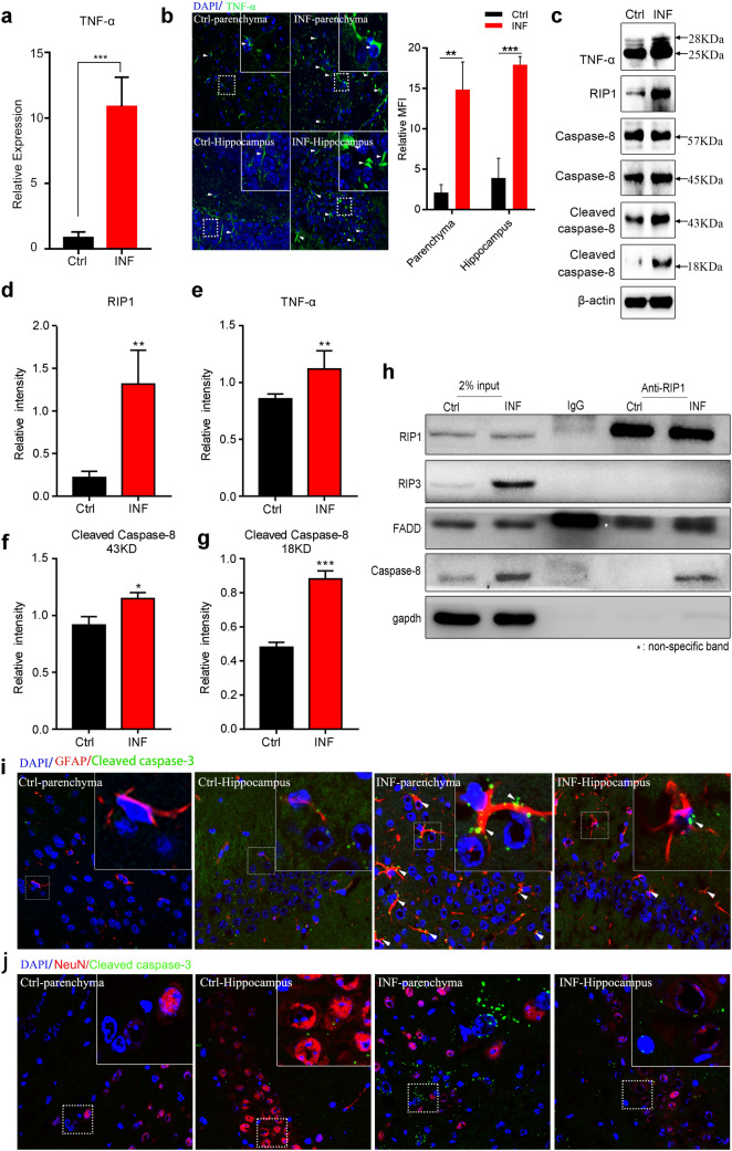
Fig. 4.
TNF-α triggers RIP1/FADD/caspase-8-mediated apoptosis of astrocytes in mouse brains with AC infection. a Relative mRNA level of TNF-α in mouse brains upon AC infection was significantly higher than that in the control group. b TNF-α protein expression level in the mouse brain post infection was remarkably elevated, as revealed by immunofluorescence (left) and relative mean fluorescence intensity (MFI, right). c The lysates of brain tissues from normal control mice or mice post infection were subjected to western blot to determine the protein levels of genes related to the apoptosis signalling pathway (TNF-α, RIP1, caspase-8 and cleaved caspase-8). d–g The protein expression levels in c were quantified via density analysis. h Mice were infected by AC for 21 days and then the lysates of mouse brain tissues were immunoprecipitated with an anti-RIP1 antibody or an IgG antibody and the precipitated complexes were separately analysed by immunoblotting with antibodies against RIP1, RIP3, FADD, caspase-8 and GAPDH. i Cleaved caspase-3 and GFAP (specific marker of astrocytes) were co-localized, as shown by immunofluorescence. j Cleaved caspase-3 and NeuN (specific marker of neurons) showed no co-localization, as shown by immunofluorescence. *p < 0.05, **p < 0.01, ***p < 0.001 (student’s t test). AC Angiostrongylus cantonensis, Ctrl normal control, INF infected by Angiostrongylus cantonensis

