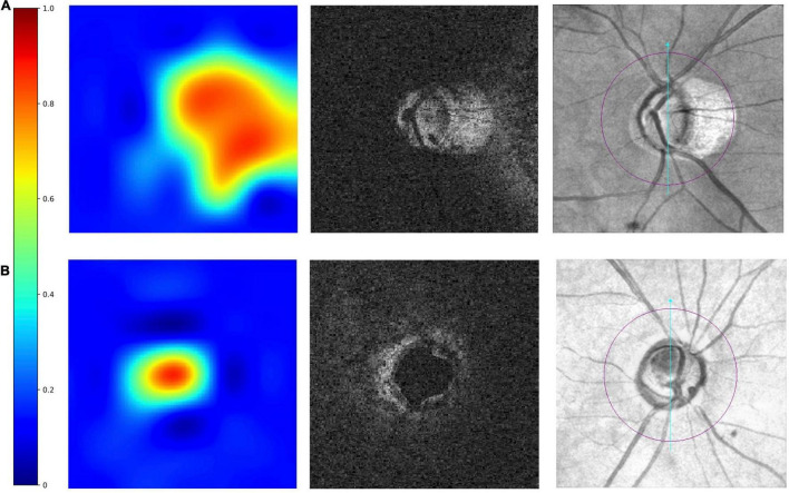
FIGURE 3.
Examples of truly detected eyes by the multi-task 3D DL model. From left to right were heatmaps, raw images, and the corresponding en face fundus images. The red–orange-coloured area on heatmaps has the most discriminatory power to detect MF. The heatmaps shows in panel (A) an eye with myopic features (peripapillary atrophy, PPA), the optic disc area and the areas with PPA is red-orange-coloured. In panel (B) an eye without myopic features, only optic disc was red-orange-coloured.

