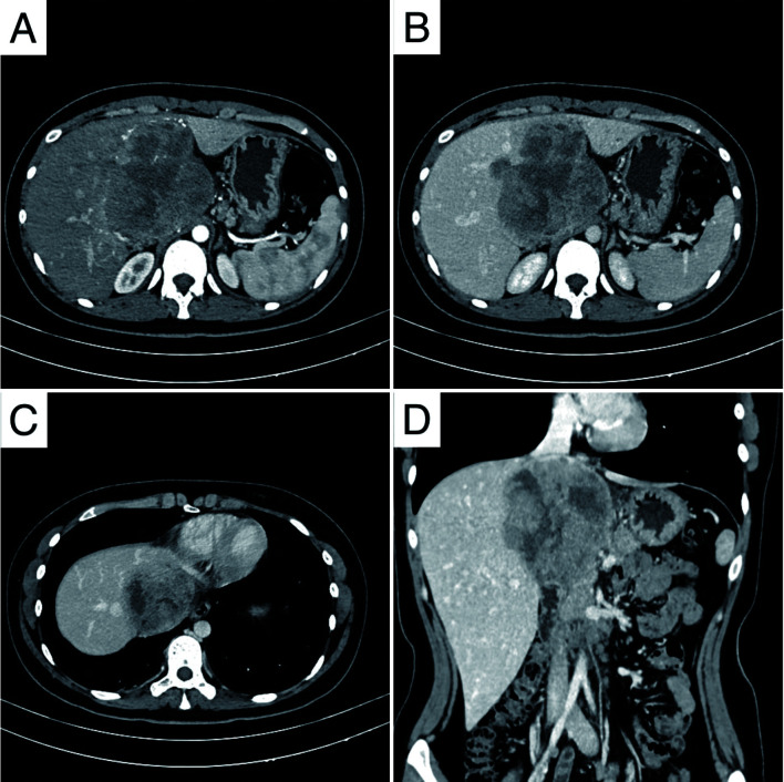
Fig. 1. Contrast-enhanced CT scans of the hepatic ES.
(A–B) Obvious heterogeneous enhancement with multiple serpentine neovascular (axial arterial phase). (C) Persistent enhancement with mild dilation of distal hepatic ducts (portal venous phase). (D) IVC involvement (coronal portal venous phase image). IVC, inferior vena cava.

