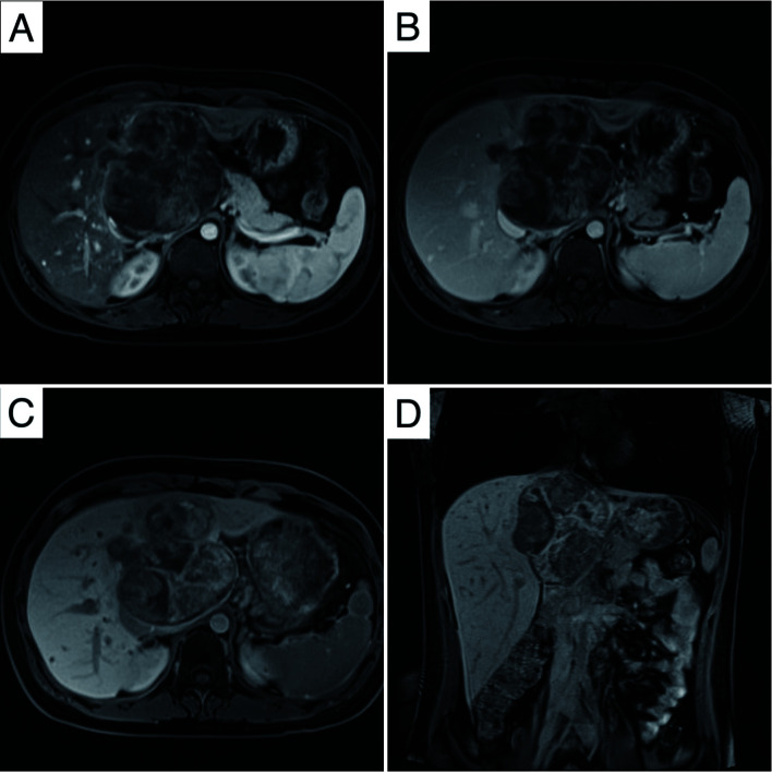
Fig. 2. Gadobenate dimeglumine-enhanced MRI of the hepatic ES.
(A) Hypointense (axial T1WI) image. (B) Heterogeneous hyperintense (axial T2WI) image. (C) Diffusion restriction in DWI. (D) IVC involvement (coronal image). T1WI, T1 weighted image; T2WI, T2 weighted image; DWI, diffusion-weighted imaging; IVC, inferior vena cava.

