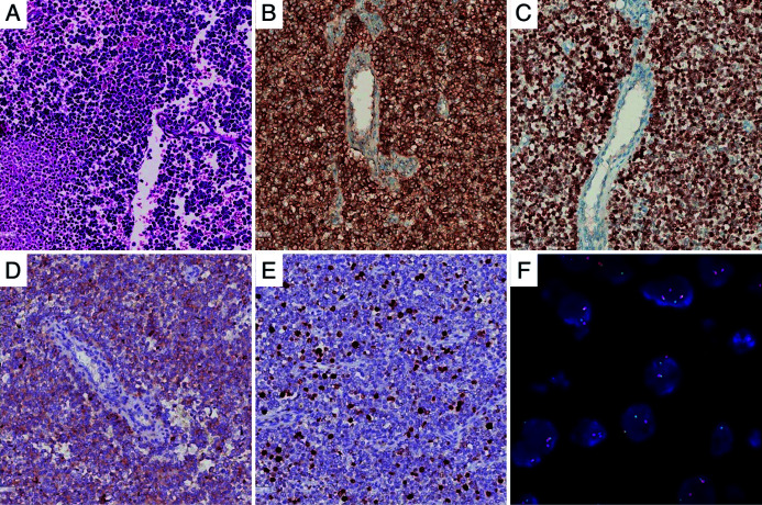
Fig. 4. Pathological and immunohistochemical staining.
(A) The tumor was composed of hypercellular small, blue-colored, round cells microscopically (HE, 200×). (B) Strong positive staining for CD 99 (IHC, 200×). (C) Strong positive staining for NKX2.2 (IHC, 200×). (D) Weak positive staining for Syn (IHC, 200×). (E) Weak positive staining for Ki-67 (IHC, 200×). (F) Dual-color, break-apart probe FISH examination, showed one yellow and one red signal, which indicated a break of the EWSR1 locus. HE, hematoxylin and eosin; CD 99, Cluster of Differentiation 99; NKX2.2, NK2 homeobox 2; IHC, immunohistochemistry; FISH, fluorescence in situ hybridization; EWSR1, Ewing sarcoma breakpoint region 1.

