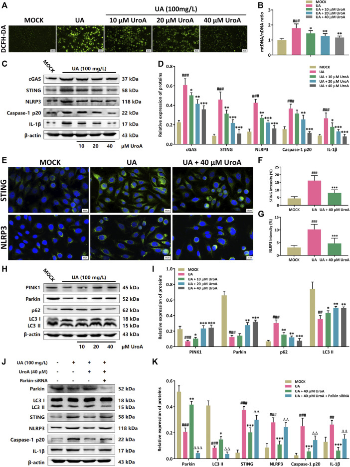
FIGURE 5.
Urolithin A (UroA) inhibits STING-NLRP3 activation by promoting Parkin-dependent mitophagy in vitro. (A) Representative immunofluorescence blots of ROS levels in the MOCK or uric acid (UA)-induced HK-2 cells with the administration of UroA (0–40 μM), respectively. (B) Mitochondrial DNA (mtDNA) to nuclear DNA (nDNA) ratio in uric acid-induced HK-2 cells with the administration of UroA. (C,D) Representative immunoblots of cGAS, STING, NLRP3, Caspase-1 p20, and IL-1β in HK-2 cells (C) and densitometric analysis (D). (E–G) Representative blots of STING and NLRP3 immunofluorescence in HK-2 cells (E) and fluorescence intensity analysis (F,G). (H,I) Representative immunoblots of PINK1, Parkin, p62, and LC3 I/II protein levels in HK-2 cells (H) and densitometric analysis (I). (J,K) Representative immunoblots of Parkin, LC3 I/II, STING, NLRP3, Caspase-1 p20, and IL-1β in the uric acid (UA)-induced HK-2 cells after parkin silencing (J) and densitometric analysis (K). β-actin was used as an internal reference. Mean ± S.D., n = 5. ## p < 0.01, ### p < 0.001 versus the MOCK group; * p < 0.05, ** p < 0.01, *** p < 0.001 versus the UA treated group; △△ p < 0.01, △△△ p < 0.001 versus the UA and UroA co-processing group.

