
FIGURE 2.
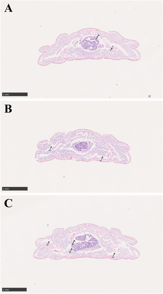
Midgut structure of P. analis. (A) Control group, (B) after 48 h of imidacloprid exposure, (C) after 72 h of imidacloprid exposure. m, midgut cell; f, normal fat body; cc, cellular compartmentation; fr, fragmentation of the midgut.
Official websites use .gov
A
.gov website belongs to an official
government organization in the United States.
Secure .gov websites use HTTPS
A lock (
) or https:// means you've safely
connected to the .gov website. Share sensitive
information only on official, secure websites.
Midgut structure of P. analis. (A) Control group, (B) after 48 h of imidacloprid exposure, (C) after 72 h of imidacloprid exposure. m, midgut cell; f, normal fat body; cc, cellular compartmentation; fr, fragmentation of the midgut.