
FIGURE 3.
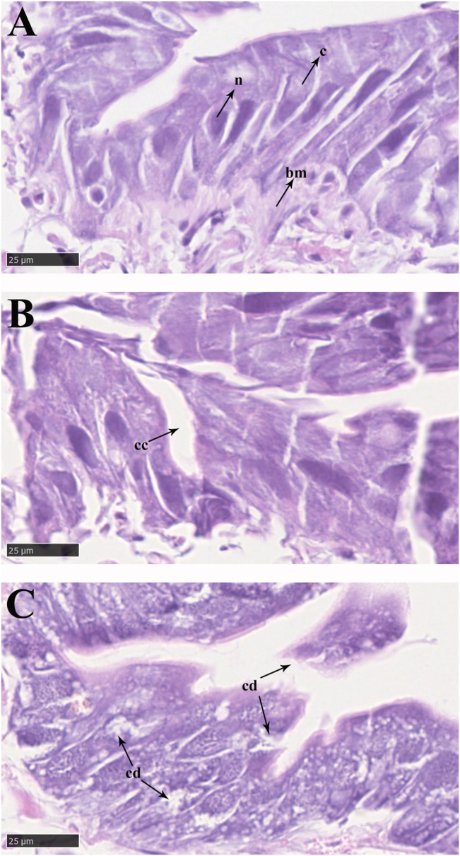
Epithelial cells in the midgut of P. analis. (A) Control group. (B) After 48 h of imidacloprid exposure. (C) After 72 h of imidacloprid exposure. bm, basement membrane; c, cytoplasm; n, nucleus; cc, cellular compartmentation; cd, cell debris.