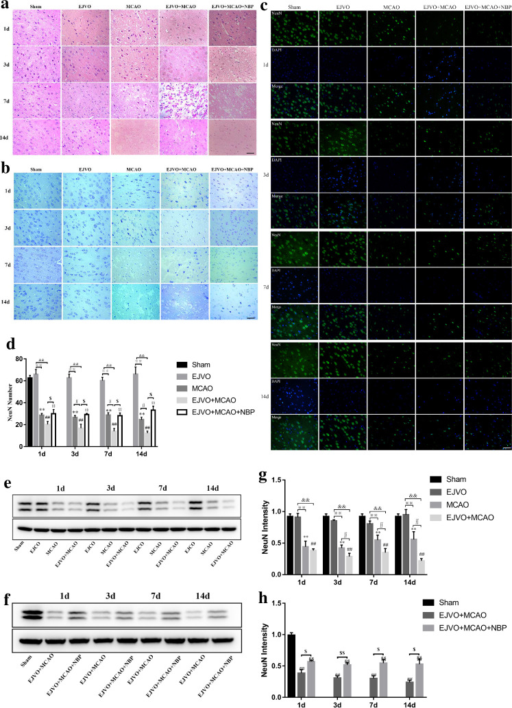
Figure 3.
Disturbance of venous circulation aggravates neuron injury after MCAO, and NBP played a neuroprotective role in rats of MCAO accompanied by EJVO. (A) Representative H&E staining showing neuron cells from first to 14th day in infarction area or corresponding area in the sham, EJVO, MCAO, EJVO +MCAO and EJVO +MCAO+ NBP groups. (B) Representative Nissl staining showing neuron cells from first to 14th day in infarction area or corresponding area in the sham, EJVO, MCAO, EJVO +MCAO and EJVO +MCAO+ NBP groups. (C) Neuron cells were labelled with NeuN antibody (green) and merged with DAPI (blue) in ischaemic area or corresponding regions of sham or EJVO group. (D) Changes of neuron cells as measured by NeuN positive cell in the different time points after operation with immunofluorescence staining. (E, F) Western blots showed NueN expression after operation treated with NBP or not. (G, H) Quantification analysis of relative expression of NeuN level. Data are presented as mean±SD, n=5 per group for immunofluorescence staining and n=3 per group for Western bolt. **P<0.01, MCAO vs Sham; ##p<0.01, EJVO +MCAO vs sham; §§p<0.01, EJVO +MCAO + NBP vs sham; ※※p<0.01, MCAO vs EJVO; &&p<0.01, EJVO +MCAO vs EJVO; ∫∫p<0.05, EJVO +MCAO vs MCAO; $p<0.05, EJVO +MCAO + NBP vs EJVO +MCAO; $$p<0.01, EJVO +MCAO + NBP vs EJVO +MCAO. Scale bar=50 µm. EJVO, external jugular vein occlusion; MACO, middle cerebral artery occlusion; NBP, Dl-3-n-butylphthalide.

